Note
Go to the end to download the full example code.
Retinal implant gallery
pulse2percept supports the following implants:
Argus Retinal Prosthesis System (Second Sight Medical Products Inc.)
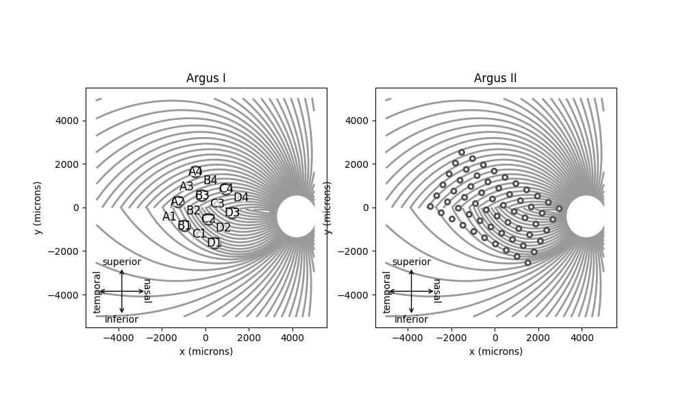
ArgusI and
ArgusII are epiretinal implants
developed at the University of Southern California (USC) and commercialized
by Second Sight. The devices were used in several clinical trials, including
NCT00279500 and NCT00407602.
Argus I is a modified cochlear implant containing 16 electrodes in a 4x4 array with a center-to-center separation of 800 um, and two electrode diameters (250 um and 500 um) arranged in a checkerboard pattern [Yue2020].
Argus II contains 60 electrodes of 225 um diameter arranged in a 6 x 10 grid (575 um center-to-center separation) [Yue2020].
import matplotlib.pyplot as plt
from pulse2percept.implants import *
from pulse2percept.models import AxonMapModel

fig, ax = plt.subplots(ncols=2, figsize=(10, 6))

# For illustrative purpose, also show the map of fiber
# bundles in the optic fiber layer:
model = AxonMapModel()
model.plot(ax=ax[0])
# Argus I is typically implanted at a 30-45deg angle:
ArgusI(rot=-30).plot(ax=ax[0], annotate=True)
ax[0].set_title('Argus I')

model.plot(ax=ax[1])
# Argus II is typically implanted at a 30-45deg angle:
ArgusII(rot=-30).plot(ax=ax[1], annotate=False)
ax[1].set_title('Argus II')
Text(0.5, 1.0, 'Argus II')
PRIMA Bionic Vision System (Pixium Vision SA)
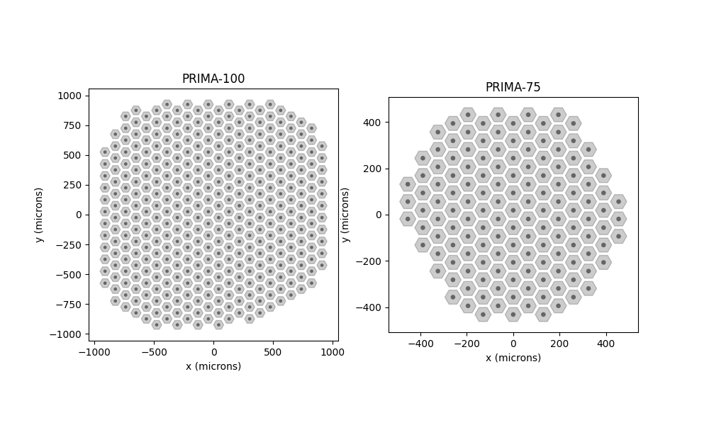
PRIMA is a subretinal device developed
at Stanford University and commercialized by Pixium Vision.
There are several versions of the PRIMA device.
The device used in clinical trial NCT03392324 consists of 378 85um-wide
pixels separated by 15um trenches (i.e., 100um pixel pitch), arranged in a
2-mm wide hexagonal pattern, and is available in pulse2percept simply as
PRIMA [Palanker2020].
PRIMA75 is a newer version of the device,
consisting of 142 70um-wide pixels separated by 5um trenches (i.e., 75um
pixel pitch), arranged in a 1-mm wide hexagonal pattern [Lorach2015].
Text(0.5, 1.0, 'PRIMA-75')
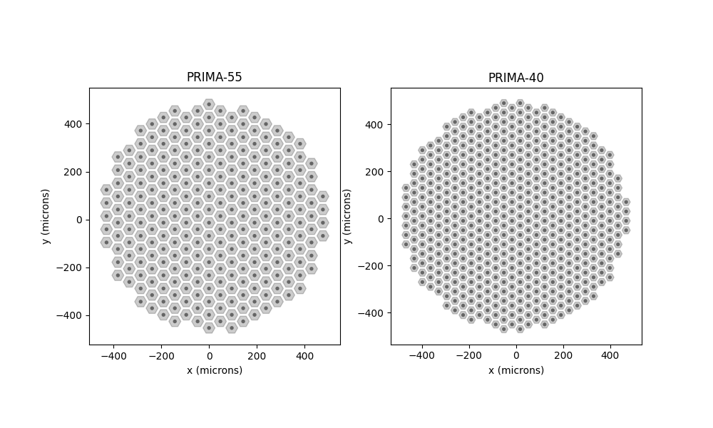
In addition, the developers are working on miniaturizing the device. At least two other prototypes are currently in development:
PRIMA55 consists of 50um-wide pixels
separated by 5um trenches (i.e., 55um pixel pitch), whereas
PRIMA40 consists of 35um-wide pixels
separated by 5um trenches (i.e., 40um pixel pitch).
The exact geometric arrangement of these two prototypes have not been published yet. The devices available in pulse2percept assume that the arrays fit on a circular 1mm-diameter substrate, which yields 273 electrodes for PRIMA-55 and 532 electrodes for PRIMA-40. These prototypes will be updated once more information about them is available.
Text(0.5, 1.0, 'PRIMA-40')
BVT Bionic Eye System (Bionic Vision Technologies)
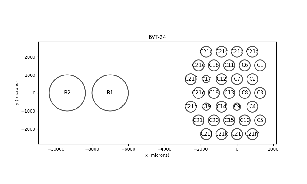
BVT24 is a 24-channel suprachoroidal
retinal prosthesis [Layton2014], which was developed by the Bionic Vision
Australia Consortium and commercialized by Bionic Vision Technologies (BVT).
Note that the array actually consists of a total of 35 electrodes:
33 platinum stimulating electrodes:
30 electrodes with 600um diameter (Electrodes 1-20 except 9, 17, 19; and Electrodes 21a-m),
3 electrodes with 400um diameter (Electrodes 9, 17, 19)
2 return electrodes with 2000um diameter (Electrodes 22, 23)
However, Electrodes 21a-m are typically ganged together to provide an external ring for common ground. Not counting the two large return electrodes leaves 24 stimulating electrodes.
fig, ax = plt.subplots(figsize=(10, 6))

BVT24().plot(ax=ax, annotate=True)
ax.set_title('BVT-24')
Text(0.5, 1.0, 'BVT-24')
Alpha-IMS and Alpha-AMS Retinal Implant System (Retina Implant AG)
AlphaIMS and
AlphaAMS are subretinal implants
developed at the University of Tuebingen, Germany and commercialized by
Retina Implant AG.
Alpha-IMS consists of 1500 50um-wide square pixels, arranged on a 39x39 rectangular grid with 72um pixel pitch [Stingl2013].
Alpha-AMS is the second generation device, consisting 1600 30um-wide round pixels, arranged on a 40x40 rectangular grid with 70um pixel pitch [Stingl2017].
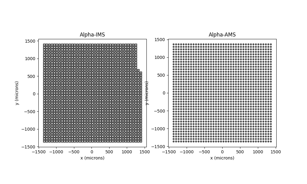
Text(0.5, 1.0, 'Alpha-AMS')
Intelligent Micro Implant Eye epiretinal prosthesis system (IMIE)
IMIE is an epiretinal implant co-developed
by Golden Eye Bionic, LLC (Pasadena CA) and IntelliMicro Medical Co., Ltd.
(Changsha, Hunan Province, China) and is manufactured by IntelliMicro.
IMIE consists of 248 large disc-shaped electrodes (210 µm in diameter) and 8 smaller disc-shaped electrodes (160 µm in diameter), arranged on an area of 4.75 mm × 6.50 mm. The center-to-center pitch is 350 µm for the large electrodes and 300 µm for the small electrodes. [Xu2021].
fig, ax = plt.subplots(figsize=(10, 6))

IMIE().plot(ax=ax, annotate=True)
ax.set_title('IMIE')
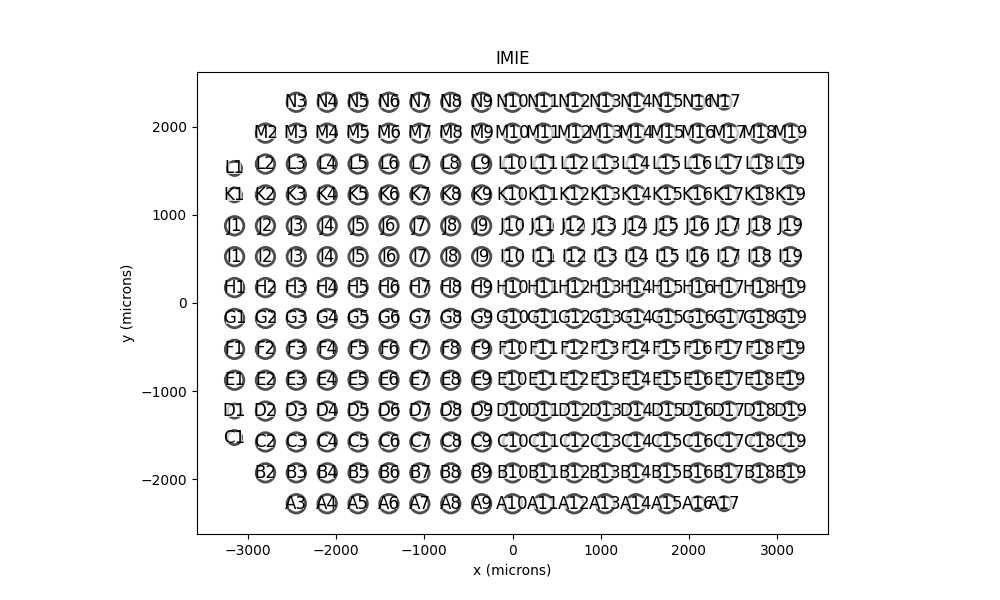
Text(0.5, 1.0, 'IMIE')
Total running time of the script: (0 minutes 1.334 seconds)